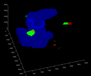
Heterochromatin in blue, E(Pc) and tou in green and red, engrailed in cyan.
These are reconstructions from confocal imaging. Still working on analysis by psf fitting and still need to do bead slide control analysis. super-resolution imaging of chromatin and these blobs of DNA at the handle and target genes should give a clearer picture of the structure at this locus.


