9:30A – 10:15 P
Fly Work
- Collect sna[18] x Double balancer virgin and male F1s
- PM virgin collection
STORM2 time.
- Colenso removing gas laser from STORM2
- Mount ultra cryo section embryo for imaging
- sections not flat or hard to find.
- remember to check filter position for quadview imaging.
For all staining reactions:
- replaced all PBS, PBT, tween and block solutions (bacteria contiminated previous block. May have contributed to reduction in antibody efficiency).
Embryo DNA FISH
- warming up post-hybe buffer for hot washes
- hot washes
- check staining — 50kbp still does not stain. Nuclear morphology otherwise looks good. Perhaps the ssDNA is titrating out the probe? Let’s try on cells in hybe buffer tonight.
- centromere embryo stain also failed. Should have done a post wash check, but really, no staining at all. This I guess renders the 50kbp stain failure meaningless. Hot wash too hot? (did have in 55C block, but didn’t warm all the way to 55C in top layer… suprised it should really all vanish too). Had used previously used 1:500 dilution, but expected it to still work.
- Repeating Cell culture stain with probe in 2x SSCT + 50% formamide (1 well, 10:200 dilution). Also a 1:600 dilution of centromere probe in same solution in 2 control wells. If this doesn’t work try a butanol free extraction?
polytene DNA FISH
- warming up 2x SSCT for hot washes
- hot wash
- restain with DAPI
- check staining
- staining looks good, took some images on confocal. Proceeded to antibody labeling
- Dm01 looks weak but detectable, preceding with H3K9me2 labeling.
- H3K9me2 antibody forze at -20C. Thawed antibody and added more sterile glycerol. Should write to millipore about this.
- Millipore says to send them the lot number and they will check. Also claim that antibody will still be good.
- Essentially no staining with H3K9me2. Weak Dm01 staining + relatively high background for normally a very clean probe. Signal looks better in smaller nuclei.
- H3-488 does label exposed polytene chromosome but does not label nuclear chromosomes, so despite methanol treatment and acetone treatment, tissue penetration is still a core problem. I guess the salvary gland tissue is made to keep things in/out. Proper mechanical rupture during prep should fix this? Maybe write and ask about staining intact polytene cells?
- Have more confocal images of polytene chromoses, should add here.
RNA FISH in situs
- warming up hybe buffer for hot washes
- hot washes
- New block arrived.
- Incubating O/N in primary antibodies
Probe making
- Take probes out of PCR, recombine into single aliquot
- Run straight on denaturing gel?
- Gel failed to polymerize. Maybe the Ammonium Persulfate has gone bad — powder was highly crystalline had to chip to get arberous fragments to dissolve. (Target volume is 300 uL as added, not 300 mL as written in protocol). Bryan provided frozen stock of 10% APS to compare it too, will try this tomorrow.
- Consider extracting without butanol, just in case that is problematic for the hybridization.
Genetic deletion screening
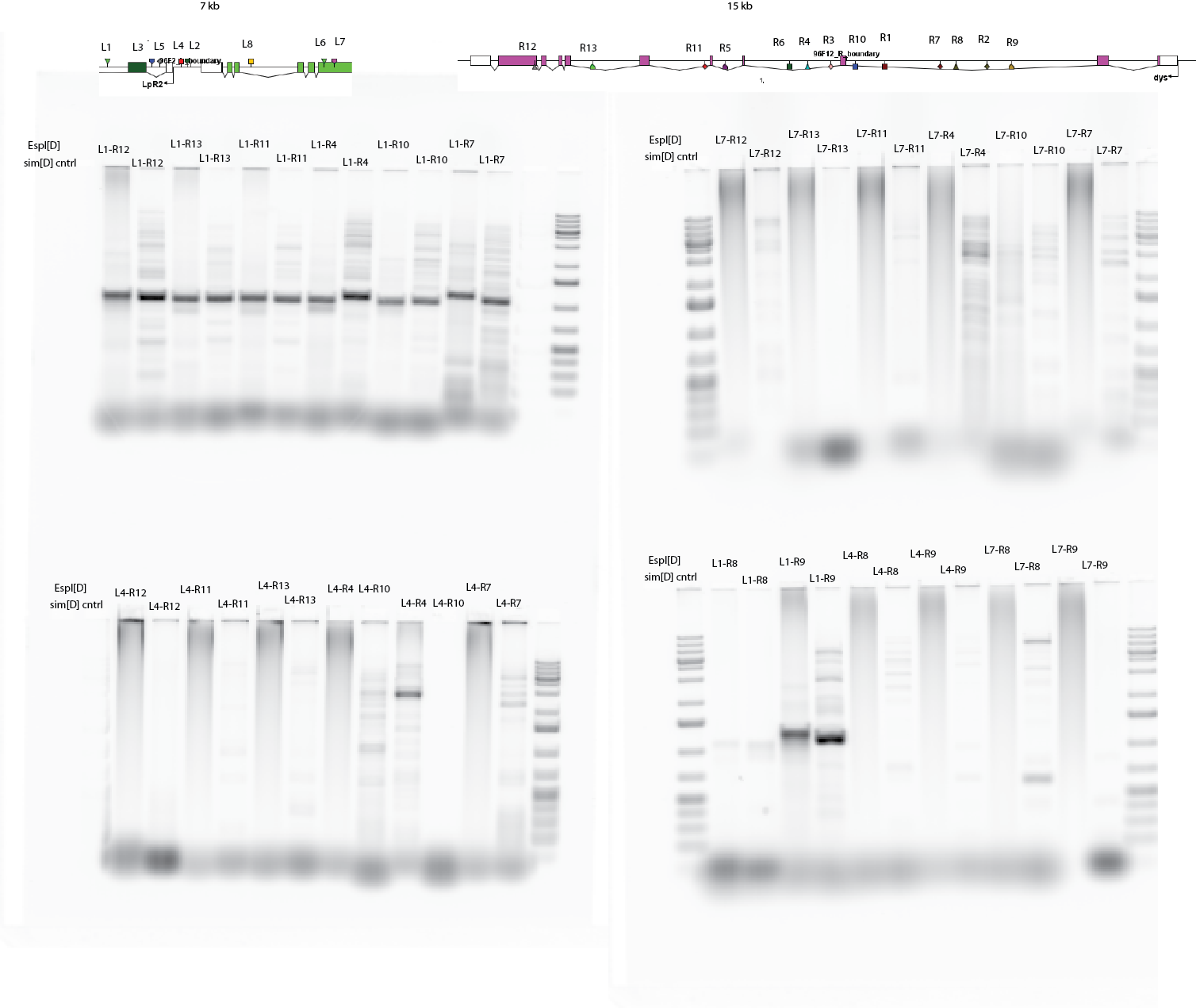
- Check Espl PCRs (48 gel lanes…)
- Strips have primers L1, L4, or L7. Each tube has primers: R12, R13, R11, R4, R10, R7, R8, R9 (moving progressively longer). Top 24 strips are Espl DNA, bottom are simD DNA.
- Run in sets of 6 pairs + 2 ladders on to separate, sequential gel runs.
- Only bands appear in sim[D] dataset. All Espl DNA lanes run as smears except with those with L1 + anything, which produce 1100 bp band from either DNA template.
- L7-R8 amplifies a 10 kb fragment in simD, L4 R4 amplifies a 3kb fragment.

- Don’t know what to do next, all 24 probe combos failed…
