Wed 07/29/15

9:10 am – 8:00 pm, 9:30 pm – 12:00 am

To do

  • confocal imaging
  • design primers
  • re-read and submit review

Done

  • confocal imaging (no staining)
  • submitted review (done)
  • feedback for Hao on essay (sent)

Embryo staining

  • no signal with 37C hybe
  • to try next: 10% formamide, 2X SCC, 10% dextran sulfate, 37C

Working on Ph co-localization data

  • see notes
  • sent updates to NJ and AW
  • analyzing Ph-M co-colocalization Flag Ph

Chromatin project

  • analyzing conventional data
  • discussed approach
Posted in Summaries | Comments Off on Wed 07/29/15

Protected: Ph-Polymerization colocalization control

This content is password protected. To view it please enter your password below:

Posted in Chromatin | Comments Off on Protected: Ph-Polymerization colocalization control

troubleshooting hb stains

Purpose

  • to start MERFISH experiments in Drosophila embryos
  • Plan: use Biosearch probes to optimize RNA labeling using DNA probes
  • currently using 20mers directly labeled. These aren’t working.

Images from Shawn Little’s paper (Gregor lab)

Gregor_Hb_images

My RNA hb stains

zoom out on whole embryo, stained in green for hb
myHb_RNA_FISH_1

a different embryo with hb in red and an intronic reporter for a hb transgene (at a different insertion site on a different chromosome) in green
myHb_RNA_FISH

My DNA hb stains don’t work at all (following protocol from the Little et al Cell paper)

  • Not clear why
  • could repeat RNA stains again just to make sure everything is working alright. Not sure if my RNA-probes are still good after 6 years in the -20C but I can try.
  • try different hybe conditions (tried RT hybe, which is what appears to be what Little et al use, tried 37C hybe).
  • could try different hybe buffers
  • I recall Raj reported not being able to get Drosophila embryos to work with his 2008 protocol
Posted in Microscopy | Comments Off on troubleshooting hb stains

Tuesday 07/28/15

9:30 am – 7:30 pm, 9:30 pm – 11:45 pm

Ph polymerization

  • reviews back
  • writing reply
  • goal to respond this week

to do

  • Ph-Flag / Ph co-localization data
  • DNA FISH long range contact frequency (hold off)

Live imaging of chromatin

  • dCAS9 for Drosophila sequence
  • transfection vector for Drosophila

Reagents

  • dCAS9-fused to Fok1 (need to add mmaple3) from addgene (here)
  • repeat sequences:
    • the “dodeca repeat” ACGGGACCAGTACGG
    • the “359 repeat” GGGATCGTTAGCACTGGTAATTAGCTGC,
    • the “AACAC” repeat: AACACAACACAACACAACACAACACAACACAACAC
    • the AATAT repeat: AATATAATATAATATAATATAATATAATAT
      DrosophilaRptLocations
Posted in Summaries | Comments Off on Tuesday 07/28/15

Monday 07/27/15

9:20 am – 10:00 pm

Reading

  • I’m looking for new insights to improve our genome wide prediction of PREs
  • Schuttengruber et al Cell Reports 2014 on PRE evolution
  • Zheng et al 2012 develop computational predictor
    • this uses ChIP data to ‘validate’ predictions
    • ChIP/damID data from Tolhuis et al (225 genes), Schwartz et al (176 genes), and Schuettengruber et al (215 genes) show only ~30 % agreement (38 genes)
    • they remove ‘duplicate genes’ from list. NO!! I want to count multiple PREs per gene as multiple PREs. don’t remove the “duplicates”, they
    • I want to see ChIP data used as input and PRE genetic tests used as validation.
    • ROC curves for this predictor outperform previous model (jPREdictor) but are still not very far off the diagonal.
  • back to Schuettengruber…
  • Schuettengruber 2014 predict 379 conserved sites within PcG domains using cross-species ChIP-seq K27me3, K4me3 (for TSS), Pc and Ph.
  • downloaded table
  • NOTE: should plot with and without peaks corresponding to TSS’s for estimated PcG density.
  • nice paper. weak, multi-component interactions specify PREs / PcG silencing, highly conserved through D. vir.
  • also has higher res Hi-C than previous Sexton et al 2012, and focuses on PcG regions.
  • claim PHO sites preferentially contact each-other uniquely in the context of PcG domains
  • show predictive correlation and KD evidence that PRC1 recruits Pho (in Ph mutants). Specifically reduce Pho binding at its PcG sites but not its non PcG domain sites.
    • mutants correlate Pho binding and Ph motif within PcG domains but wt does not (supporting cooperative recruitment model).
    • outside of PcG context, Pho co-localizes with CP190 and BEAF32

Review

  • finish refereeing paper and submit recommendations (due tomorrow)

Chromatin project

for analysis of deviants, comparison to other features

  • getting D mel embryonic gtf file to run cufflinks
  • SAM format specs
  • SAM files need to be sorted first by SAMtools see thread.
  • data is not presorted the way cufflinks needs, (should be alpha-numeric by chromosome).
    SAMfileNotSorted
  • sorting using this command:
    sort -k 3,3 -k 4,4n hits.sam > hits.sam.sorted
  • (From BioSTAR: The code just means to sort on column 3, then by column 4(numerically) of the hits.sam file and print to hits.sam.sorted)
    e.g.
    sort -k 3,3 -k 4,4n /n/home05/boettiger/Genomics/Data/GSE18040_Dm_KC167.sam > /n/home05/boettiger/Genomics/Data/Dm_Kc167.sam.sorted 2> /n/home05/boettiger/Genomics/Data/errorsSort_KC167.txt
  • best to test these things on small data sets
  • wrote matlab command to build smaller dataset (ParseSAMdata_150727.m)
  • sort command works as expected on this.
  • this runs properly through cufflinks (tested small version)
  • sorting the whole 3Gb data set on Odyssey with this command is very slow…
  • sorting finished, running cufflinks still failed. Upset about ordering of chr M and chr U in the SAM file (neither of which I need!!)
    • Moreover this file IS sorted correctly, U is after M (and before X and Y) so shut up and keep analyzing!
    • 'current' 'hit' 'is' 'at' 'U:3652,' 'last' 'one' 'was' 'at'
      'M:18987'
  • samtools sort doesn’t work on sam files, only bam files (so much for “sam”tools).
  • file REFUSES to convert to BAM becuase there is no @SQ lines in the header.
  • okay, so let’s sort by hand and remove the ‘M’s and ‘U’s using matlab
  • matlab textscan reads this into inefficient cell arrays, which are now using ~60 Gb (yes gb) just to textscan in a ~4 Gb text file.
  • Bogdan is going to fix this in Python
  • after some more frustration, data ran correctly.

RNAi

qPCR

  • setting up qPCR of last weeks PPPES (1 and 2) and Ph KD samples, along with corresponding mocks.
  • assay for 3 cntrl genes, 3 PcG targets, + Pc and Ph-p.
  • column order (cDNA): PPPES 1, PPPES2, Ph-Kd, Ph-Kd-mock, PPPES-mock, prior-mock
  • row order (primers): alpha-tub, act, gapdh, Pc, Ph-p, Antp, Abd-B, en
  • flipped primer labels. oops. fortunately I sorted by expression so it’s easy to spot.
    qPCR_plate_150727 qPCR_plate_CI_bounds_150727

Embryo staining

  • check samples on confocal
  • no staining at all.
  • maybe 37 C is necessary.
  • previous results look vaguely more encouraging
    NoStainingEarly NoStainingEarly_Q RNA_FISH_with_DNAprobes_1 RNA_FISH_with_DNAprobes_overlay

Issues with protocol

  • Temperature not mentioned. I assume this means RT but I find that a bit strange for hybes
  • probe sequences, probe length, and probe number not mentioned

Other

  • gave 8 uL of 40ng/uL YW gDNA to AC.
Posted in Summaries | Comments Off on Monday 07/27/15

Sunday 07/26/15

3:30 pm – 6:30 pm

embryo stains

  • rinse embryos in pre-hybe/ was buffer, 1 hr
  • rinse embryos in PBT
  • stain a subset with WGA
  • mount this subset for confocal imaging in Prolong Diamond

Chromatin project

  • discussion on project progress with BB
  • email CW about antibody stains.
Posted in Summaries | Comments Off on Sunday 07/26/15

Friday 07/24/15

9:15 am – 7:15 pm

PRE mapping

  • Ringrose 2003 predicted PREs http://www.techfak.uni-bielefeld.de/~marc/pre/hit_list.html
  • Ringrose 2003 is dm1. Need to lift over to dm3 before we can map.
  • lift over, plotted correlation.
  • serious computer crash (windows froze while unpacking .gz file), matlab history corrupted,
  • not too strong a correlation with the motif derived ChIPseq data.
    rg_vs_ringrosePREsvolume_vs_ringrosePREs
  • using peaks from Follmer and Francis data
  • these peaks look less PcG specific, the ones in the PcG’s match the ones I’d infere from modENCODE
    en_S2cells_K27me3_Psc_Pc
  • here’s the correlation:
    rg_vs_follmerPeaks volume_vs_follmerPeaks
  • should comment on pairing dependent silencing and (more importantly) multiple insertion silencing (see Kassis 2013 review).

Cell Prep

  • poly-Lysine coated 24 coverslips in 12 well chambers
  • plated Ph-p Ph-d RNAi KD cells onto coverglass 10 per plate + 2 WT/mock controls
  • fixed cells, now in PBS in 4C

RNA isolation

  • 2 1.5 mL eppendorfs each
  • ran the Qiagen RNeasy extraction
  • high concentrations this time (Ph ~1600 ng/uL and mock at 1400 ng/uL)

Maxima cDNA first strand synthesis

  • protocol
  • per reaction
    • (2x the recommended mix for a 40 uL reaction)
    • 3 uL 100 uM primer (= 300 pmol)
    • 3 uL 10 mM dNTPs
    • 10 uL RNA
    • 14 uL ddH2O
    • heat to 65C for 5 min
  • then add per reaction
    • 8 uL 5x RT buffer
    • 2 uL enzyme
    • 2 uL RNasin
    • mix, run at 50C 1 hr, then heat denature.
  • froze extra RNA at -80C
  • cDNA reaction still running.

Ordering

  • ordered finer forceps (I the cheap ones I bought are too stiff and I keep dropping my coverslips)
  • ordered more coverslips
Posted in Summaries | Comments Off on Friday 07/24/15

Thursday 07/23/15

12:30 pm – 7:00 pm

(sick)

RNAi experiments

Primer issues

  • diagnosing problems with primers for qPCR
  • writing code for primer mapping
  • organizing a better data structure to keep track of all the RNAi targets, their qPCR primer sets, their RNAi primer sets, their accession numbers, etc.
  • my overlap checking clearly did not work correctly. Overlap of qPCR and RNAi primers detected for RNA
  • additionally, several of the 20mer primers have perfect hits elsewhere in the genome. Only one should have a sufficiently close compliment to be able to amplify so it shouldn’t be a problem, but still, would have been good to check sooner
    qPCRandRNAiPrimerLocations
  • note Pc_v1, Esc, Pho, and Su(z)2 qPCR primers all overlap RNAi
  • and apparently the polyA selection for cDNA conversion does not sufficiently screen out the added RNA

RNAi new knockdown

  • 2 6-well wells with Ph-p + Ph-D (30 mg/mL)
  • 4 mocks
  • used cells from SFX, rinsed in PBS and repelleted
  • one 75mm2 flask, could have used a few more cells for a bit better density, but it is okay.
  • incubated 55 min.

Cell culture

  • passaged Psc- cells with Trypsin.
  • washed cells with PBS to remove excess serum (inhibits Trypsin)
  • added 3 mL Trypsin soln (now in 4C).
  • After 20 min, they blow off easily. (After 10 they just start rounding up. Next time try just 10 min)
  • add a bit of media to try and inhibit trypsin and pellet cells
  • wash in 5 mL PBS (resuspend and repellet)
  • resuspend and plate in 75mm flask in fresh media.

Embryo in situs

  • remade hybe buffer, dissolving BSA in H2O first (never went into solution otherwise).
  • hydrating embryos into PBT
  • diluted probes (5 nmol) to ~50 uM in ddH20, 50 probes so individual probes at 1 uM. recommended final concentration at 1 nM per probe. Lets try 2 uL in 20 uL hybe buffer O/N. Thats ~10 nM.
Posted in Summaries | Comments Off on Thursday 07/23/15

Wednesday 07/22/15

9:30 am – 8:45 pm

Psc deletion

  • picked up psc deletion cells from Sonny, along with media
  • (also gifted a second aliquot of the Pc-antibody), now frozen.
  • also gifted ~200 mL of media ready mixed.
  • cells were last plated 7/12 and passaged 1/4. Currently ~60% confluent.
  • should remove with trypsin, need to spin down, wash, spin down again, and resuspend to passage.

RNAi experiments

Imaging

  • washed cells, 40 min at 60C in 2x SSC
  • added 100 nm 540/560 beads at 1:3000 (this density looks great)
  • imaging cells – nice and bright, decent density, good contrast. Hopefully these STORM well.
  • the TIRF on STORM2 is all kinds of weird
  • started new AlistairTemp (2 Tb) for data collection on STORM2 (eSATA is functional)
  • installed gitk on storm2 computer
  • copy paste of storm2_splitdax imagewriters.py into latest storm2 branch appears to work fine.
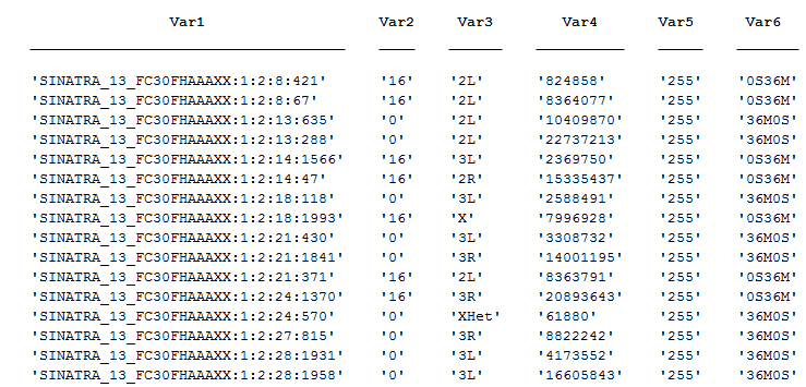
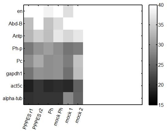
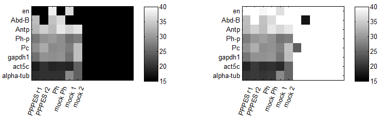
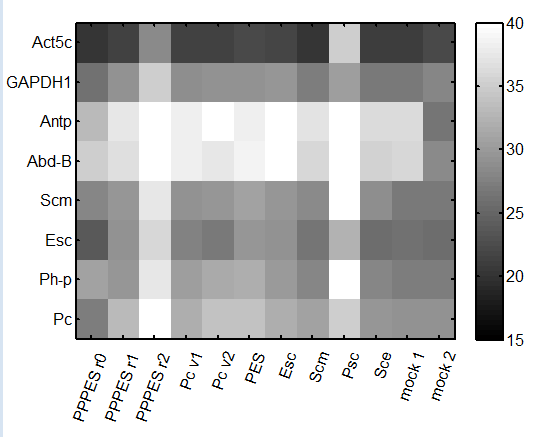
repeat qPCR

  • try lower concentration of cDNA – 1 uL per reaction
  • primers: Pc, Ph, Abd-B, en, Antp, alpha-tub84, Act5A, gapdh1
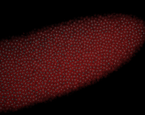
embryo FISH

protocol from Little 2013

“Embryos were fixed in5%formaldehyde, 1XPBSfor 20 min, and devitellinated as described by Lecuyer et al. (2008). Fixed embryos were rinsed three times in 1X PBS and washed for 10 min in smFISH wash buffer (4X SSC, 35% formamide, 0.1% Tween 20). Hybridization to probes complementary to the reading frame of hb, Kr, kni,or gt and conjugated to Atto 565 (Sigma-Aldrich; 72464) or Atto 633 (Sigma-Aldrich; 01464) was performed for 16–24 hr at a concentration of about 1nMper probe in hybridization buffer (4X SSC,35%formamide,10% dextran sulfate, 2 mg/ml BSA [NEB; B9001], 0.1 mg/ml salmon sperm DNA [In- vitrogen; 15632-011], and 2 mM ribonucleoside vanadyl complex [NEB; S1402S], 0.1% Tween 20). After two washes of 1 hr in wash buffer, embryos were rinsed twice briefly in 1X PBS, stained with DAPI, and mounted in VECTA- SHIELD (Vector Laboratories; H-1000)”

buffers

  • prehybe buffer: 4X SSC, 35% formamide, 0.1% Tween 20 (50 mL master)
    • 10 mL 20x SSC
    • 17.5 mL formamide
    • 500 uL 10% Tween 20
    • fill to 50 mL with ddH2O
  • hybe dilution buffer: 4X SSC,35%formamide,10% dextran sulfate, 2 mg/ml BSA, 0.1 mg/ml salmon sperm DNA, and 2 mM ribonucleoside vanadyl complex , 0.1% Tween 20 (50 mL master)
    • 10 mL 20x SSC
    • 17.5 mL formamide
    • 10 mL 50% dextran sulfate
    • 100 mg BSA
    • 500 uL of 10 mg/mL sonicated salmon sperm DNA
    • RVC
    • 500 uL 10% Tween 20
  • would have been better to pre-disolve the BSA in 10 mL of ddH2O — it does not want to go into solution afterwards. heating to 37, will rock O/N.

Chromatin paper

  • discuss comments with XZ and BB
  • need action items for XZ asap.
Posted in Summaries | Comments Off on Wednesday 07/22/15

Tuesday 07/21/15

10:15 am – 8:00 pm

RNAi experiments

Quantifying knockdown efficiencies by qPCR

cDNA

  1. PPPES (Pc, Ph-p, ph-d, Esc, Scm) rep 1
  2. PPPES (Pc, Ph-p, ph-d, Esc, Scm) rep 2
  3. Pc v1
  4. Pc v2
  5. Pc, Esc, Scm,
  6. Esc
  7. Suz12
  8. Scm
  9. SCE
  10. mock
  11. PPPES (Pc, Ph-p, ph-d, Esc, Scm) rep 0 (from last week)

genes to test

  1. Pc
  2. Ph-p
  3. Esc
  4. Scm
  5. Suz12
  6. SCE
  7. Abd-B
  8. Antp
  9. Ph-d
  10. Gapdh1
  11. Act5A

Experiment layout

  • Note: don’t have primers for Su(Z)12
  • see qPCR setup google doc
  • 2.5 uL of primer mix
  • 2 uL of cDNA

Results

  • knockdown doesn’t seem to have worked so well this time.
  • maybe some issues with the qPCR, will try to run a few lanes again.
    qPCR_plate_150721
  • Notes
    • col 9 has Psc instead of Scm
    • col 10 has Sce instead of Scm
    • col 12 has ‘Psc, Sce, Ph-D and alpha-tub84a’ instead of ‘Abd-B, Antp, Gapdh1, and Act5c’ respectively

new dsRNA synthesis

  • freeze T7 reactions of original PPES combo. Not sure we’re coming back to this.

STORM

  • cells very sparse.
  • note: the top filter wheel sometimes gets turned. It needs to be in position 1. take off the inspection port. if empty port 3 is visible in the inspection port, the correct port 1 is aligned. The IR focus lock beam bounces off this dichroic. No idea why people swap this out.

New stains

  • prepped whole plate of cells
  • should make initial density lower still — a lot of cells detach in sheets during the LN2 treatment.
  • seems to me the PcG RNAi treated cells detach more than the wt / mock cells do.
  • RNase treated just 2 coverslips. Rest still RNA-intact.
Posted in Summaries | Comments Off on Tuesday 07/21/15